Ref: http://www.ncbi.nlm.nih.gov/pmc/articles/PMC3134027/
O3 Ozone Wound Healing - Images
1. Case Study 01:
The following images are medical records of an infected tibia and knee joint after surgery.
This was a non-healing wound. After 15 days of antibiotic treatments with no response ... amputation was recommended to the patient.
The patient sort a second opinion from an oxygen medicine doctor ...
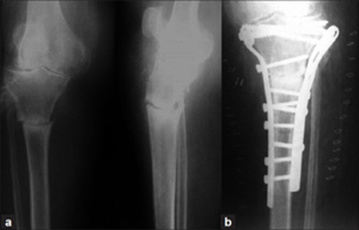
Fig. 1. (a) Radiograph showing stress fracture of proximal tibia; (b) Dual plating using two incisions as the fracture did not show any signs of healing with 1 month of plaster cast.

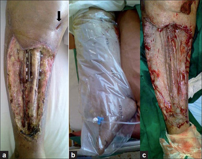
Fig 2. (a) Clinical appearance of the wound at the time of presentation showing necrotic tissue and exposed bone and draining wound communicating to the knee joint (arrow);
(b) Limb being exposed to topical ozone therapy;
(c) Split thickness skin graft on the wound.

2(a) The wound which would not heal after surgery and 15 days of antibiotic drug medication.
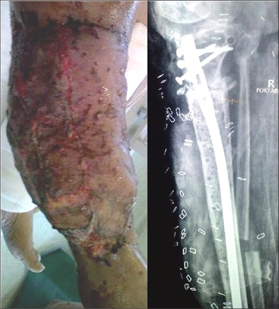
Fig 3. (a) Latissimus dorsi pedicle flap was done to cover the wound and implant removal and intramedullary nailing was performed in a single stage.

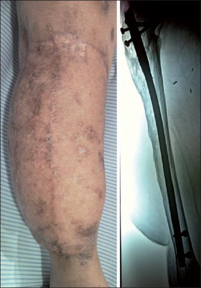
Fig 4. (a) Clinical and radiographic appearance at 20 months postsurgery showing healing of both soft tissue and the bone.

________________________________________ |
|